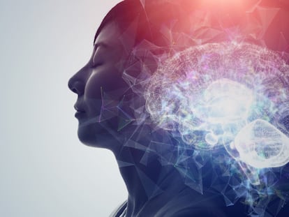
Experimental research on heading perception: therapeutic applications and their benefits
As you progress through the environment, moving images form on your retina. This “optic flow” originates from your – the observer’s – navigation. This enables you to perceive and interact with your surroundings. Computing the correct heading may be a very simple brain function for straightforward objectives – like reaching the top of a flight of stairs – but in everyday life self-motion perception is a very complex function, requiring the integration of our own movements with all the objects, people, animals, etc., in the vicinity.
Persons with cortical damage, which may be caused by cerebral strokes, are often unable to navigate successfully in the environment. This is mainly caused by the inactivation of neuronal circuits linking the various areas involved in heading perception and movement generation. Our neurophysiological studies performed on primates, at both the University of Bologna, Italy and at Rutgers University of Newark, New Jersey, USA, provide a new perspective on potential rehabilitation therapies for some patients suffering from these difficulties. Primate research is a powerful approach that allows accurate characterization of normal cortical functions. Such research provides crucial information on brain function that cannot be obtained in any other way.
The cortical areas of the brain seem to be involved in optic flow processing – for example, allowing one to perceive the correct heading. Evidence suggests that such processing in action evolves across different cortical areas. Specific circuits in the cortical area are likely to be responsible for successive modifications in the way the optic flow is represented within the brain. Each cortical area utilizes a unique combination of signals. These combinations require a specific connectivity between not just pairs of cortical areas, or pairs of neurons, but also between specific parts of each cortical representation. When a cortical area is damaged, the ability to integrate visual signals with other sensory inputs fails.
Although much is known about the cortical representation of the optic flow on the retina, the mechanisms underlying signal processing are largely unknown and the manner in which these cortical areas contribute to actual perception remains an open question. In answering this question, particular attention has to be paid to the specific cortical connections of each area and to the activity of target neurons. Recently, a novel concept was proposed: that no hierarchy is necessary in the utilization of optic flow and that each cortical area forms a stable representation of the information appropriate to its particular function. In both the early visual system and late motor system the representation of cortical structures and functions – the so-called topography – is of the utmost importance, providing a better understanding of the connectivity between areas of the brain and the self-organizing principles of cortical development.
In order to uncover the cortical topography, we study the activation of brain areas while the animal performs a specific task, such as fixating a little spot during the presentation of visual stimuli. These results can then be transferred to human subjects. Our research on primates has recently revealed the cortical topography of the optic flow representation in a specific region of the brain known as the parietal cortex. Our hypothesis is that such topography might be crucial to perceive heading direction when the observer does not fixate the final destination of his/her motion. Another issue also appears to be very important: the topographical representations were not stationary over a period of days, suggesting that they are plastic – it appears that neurons are constantly modulated to provide a short-lived, but useful, representation of the optic flow.
Neurological patients with parietal lobe lesions may benefit from greater understanding through the development of improved disease treatment, leading to better rehabilitation therapies. Studies of this kind – performed on primates – are crucial for determining which parietal representations are important to focus on for modification by therapy when there are spatial deficits in human subjects caused by parietal damage. The next step is to broaden our knowledge of the neural mechanisms underlying heading perception processing and how damage in parietal areas can impair specific navigation features. This kind of research has great potential to make a lasting contribution to neurological sciences, since it provides insight into a brain function that is fundamental to cognition, namely selective attention, which is our ability to choose what we want to perceive and act upon.
Tu suscripción se está usando en otro dispositivo
¿Quieres añadir otro usuario a tu suscripción?
Si continúas leyendo en este dispositivo, no se podrá leer en el otro.
FlechaTu suscripción se está usando en otro dispositivo y solo puedes acceder a EL PAÍS desde un dispositivo a la vez.
Si quieres compartir tu cuenta, cambia tu suscripción a la modalidad Premium, así podrás añadir otro usuario. Cada uno accederá con su propia cuenta de email, lo que os permitirá personalizar vuestra experiencia en EL PAÍS.
¿Tienes una suscripción de empresa? Accede aquí para contratar más cuentas.
En el caso de no saber quién está usando tu cuenta, te recomendamos cambiar tu contraseña aquí.
Si decides continuar compartiendo tu cuenta, este mensaje se mostrará en tu dispositivo y en el de la otra persona que está usando tu cuenta de forma indefinida, afectando a tu experiencia de lectura. Puedes consultar aquí los términos y condiciones de la suscripción digital.